Key points#
- Automated, AI-based systems can analyze retinal photographs and flag referable diabetic retinopathy, and some are authorized for use in settings such as primary care.
- A program may expand screening reach, but benefit depends on eligible patients, usable images, referral capacity, completed follow-up, and effective treatment when indicated.
- Their scope is narrow. They are built to detect referable retinopathy, not to replace a full eye examination that can find other conditions.
- They depend on good-quality images and on clear pathways to specialist care for positive results, so human oversight still matters.
Few areas generate more noise than artificial intelligence in medicine, and eye screening is one where the technology is genuinely real and already in use. That makes it a good place to practice reading these stories honestly, somewhere between the breathless headlines and the reflexive dismissal. The most useful question is not whether AI eye screening is good or bad, but what it actually does, where it helps, and what it cannot do.
What these systems actually do#
Diabetic retinopathy is assessed by looking at the retina, and retinal photographs capture that view. Automated systems analyze those photographs within a defined intended use. For gradable images, a system may return a bounded result such as below a referral threshold or referral indicated; an ungradable or out-of-scope image still needs a reviewed next step. The output is not a complete diagnosis or eye examination.
This is a narrow and well-defined task, which is exactly why software can do it. Some of these systems have been authorized for use in everyday settings such as primary care offices, where a photograph can be taken and assessed without a specialist physically present. Guidelines for diabetes care recognize validated retinal photography, including approaches that use automated interpretation, as an acceptable way to screen.
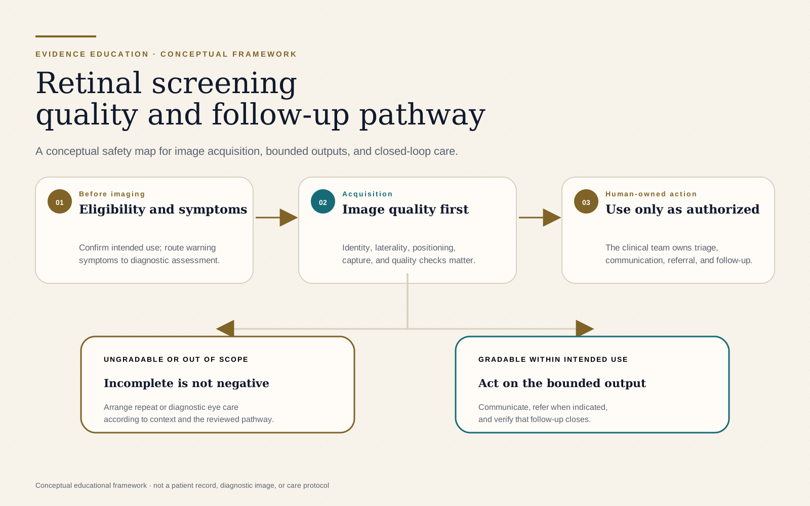
Scroll horizontally to inspect the full figure. A complete text version follows.
Read the figure in text
Before imaging, confirm intended use and route warning symptoms to diagnostic assessment. During acquisition, verify identity, laterality, positioning, capture, and image quality. Apply an output only within the device's authorized intended use; the responsible clinical team owns triage, communication, referral, and follow-up. Ungradable or out-of-scope results lead to repeat or diagnostic eye care.
Where it genuinely helps#
The potential program-level benefit is not that software is cleverer than a specialist. It is that screening may be offered in more settings. Many people with diabetes do not receive eye screening as often as recommended, and diabetic retinopathy can remain silent while it progresses.
Bringing screening into primary care, community clinics, or settings far from specialist eye care may reduce one access barrier. That possibility is not automatic benefit. The program still needs eligible patients, usable images, prompt communication, referral capacity, completed follow-up, and treatment that improves outcomes. If any link fails, an accurate screening output may not help the person screened.
The limits worth understanding#
An honest account has to include the boundaries, and they are important.
- Image quality matters. These systems depend on getting a good photograph. Poor images can mean a result the system cannot interpret, which then needs a human pathway.
- The scope is narrow. A tool built to detect referable diabetic retinopathy is not looking for everything. A complete eye examination can find other conditions, from glaucoma to other retinal problems, that a focused screening tool is not designed to detect. Screening is not the same as a full eye exam.
- A positive result needs somewhere to go. Flagging disease only helps if there is a clear, reliable route to specialist assessment and treatment. The technology is one link in a chain, and the chain has to be complete.
- Oversight remains. Validated performance on the specific task does not remove the need for clinical judgment, sensible follow-up, and attention to the individual patient.
None of these limits is a reason to dismiss the tools. They are the reasons to use them well, as part of a thought-through pathway rather than a standalone answer.
How to read claims about it#
This topic is a useful template for reading any claim about AI in medicine. Ask what specific task the system performs, how well it has been validated for that task, what its scope does not include, and what happens to the people it flags. A system can be genuinely good at a narrow job and still be oversold if those boundaries are blurred. Holding both ideas at once, real usefulness and real limits, is the honest position.
Where this leaves patients and clinics#
An automated retinal screening offered during a primary care or community visit can be a useful focused step for an eligible person, especially when access to eye care is difficult. It is not a complete eye examination, and its value depends on image quality, intended use, communication, referral, follow-up, and access to effective care. A clinic should be able to explain what each possible output means and who owns the next action.
Sources and further reading
Questions and answers
Can AI screen for diabetic eye disease?
Yes, within a defined intended use. Authorized systems can analyze retinal photographs and return a bounded screening result that may indicate referral, repeat imaging, or another reviewed next step. They do not replace a complete eye examination or make every diagnosis.
Is AI screening as good as a specialist?
For the specific task of detecting referable diabetic retinopathy from good-quality images, validated systems can perform well. But their scope is narrow. They are built to catch one thing, and they do not replace a complete eye exam, which can find other conditions a screening tool is not designed to detect.
What is the main benefit of AI eye screening?
A well-run program may expand screening reach. Benefit still depends on selecting eligible patients, obtaining usable images, communicating results, having referral capacity, completing follow-up, and providing effective treatment when indicated.
What are the limits?
The systems need good-quality images, they focus on referable retinopathy rather than all eye disease, and a positive result still needs a clear pathway to specialist care. Human oversight and sensible follow-up remain essential.